Arthroscopy
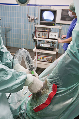
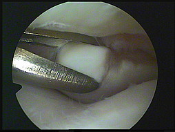
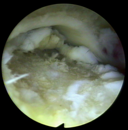
Arthroscopy enables us to take a view at the inside of a joint on a monitor. Arthroscopy is a minimally invasive and thus very gentle procedure. Through an incision of app. 1cm in length, an arthroscope is introduced into the joint. Surgical procedures can be carried out through a second incision which is also minimal.
Arthroscopy is used both diagnostically and for treatment. Diagnostically, it is particularly useful for the exact assessment of the degree of damage such as cartilage defects or synovial villi disorders and for issuing a more accurate prognosis.
Most commonly, arthroscopy is carried out to remove OCD (Osteochondrosis dissecans) or bone fragments respectively from the joint. Particularly in the coffin joint, pastern joint, fetlock joint, stifle and tarsal joint, “joint mice” or bone chips are common. These bone chips can act like sand in the gears and cause acute or subsequent lameness. Preventive removal of a bone fragment can also make sense.
During arthroscopy, cartilage defects can be smoothed out and meniscal defects in the equine stifle (e.g. meniscal tears) can be surgically treated. Additionally, arthroscopy allows the option of flushing infected joints. In cases of open joint injuries, foreign bodies and contaminations can be removed under visual control. In foals with joint-ill, arthroscopic flushing of the joint is also the method of choice to flush out bacteria.
Treatment of bone cysts is a further important field of application of arthroscopy.
![[Translate to English:] Kleintiermedizin Hochmoor [Translate to English:] Kleintiermedizin Hochmoor](/kv/_processed_/7/e/csm_Kleintiermedizin-Hochmoor_c796a9e531.jpg)
![[Translate to English:] Klinik für Pferde [Translate to English:] Klinik für Pferde](/kv/_processed_/0/d/csm_pferdeklinik-01_c18cc541af.jpg)
![[Translate to English:] Allgemeine Untersuchung Kleintier [Translate to English:] Allgemeine Untersuchung Kleintier](/kv/_processed_/1/5/csm_allgemeine-Untersuchung-Kleintier_cdba7ebb98.jpg)
![[Translate to English:] Lahmheitsuntersuchung Pferdeklinik Hochmoor [Translate to English:] Lahmheitsuntersuchung Pferdeklinik Hochmoor](/kv/_processed_/2/e/csm_Lahmheitsuntersuchung-Pferdeklinik-Hochmoor_8e9ec18025.jpg)

![[Translate to English:] Tiermedizin Hochmoor CT Hund [Translate to English:] Tiermedizin Hochmoor CT Hund](/kv/_processed_/6/4/csm_Kleintiermedizin-3_56aa5d056b.jpg)
![[Translate to English:] Tiermedizin Hochmoor Kleintiermedizin [Translate to English:] Tiermedizin Hochmoor Kleintiermedizin](/kv/_processed_/d/e/csm_Tiermedizin-Hochmoor-Kleintiere_5f8df77a2f.jpg)
![[Translate to English:] Pferdemedizin Hochmoor [Translate to English:] Pferdemedizin Hochmoor](/kv/_processed_/5/6/csm_Pferdemedizin-Hochmoor_2a06563c44.jpg)

![[Translate to English:] Gelenkchips auf dem Röntgenbild Bone chips on a radiograph](/kv/_processed_/8/b/csm_Neues_Bild2_70b4192c6b.jpg)

