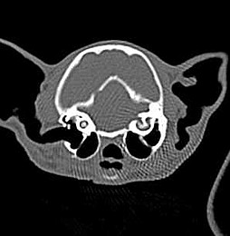
Computertomography
Computed tomography is an advanced diagnostic method which works with X-radiation and is usually applied when a certain part of the body or a limb has to be examined more closely.
For example, CT examination can be helpful in horses where the area causing lameness has been identified using nerve blocks, but a diagnosis could not be reached on clinical examination, normal X-rays and ultrasonography. The head is also an area where an exact diagnosis is often only possible after imaging of the anatomical structures without superimposition during computed tomography.
Contrary to conventional radiography, X-rays are not projected onto radiographic film during computed tomography but are collected by a detector which measures radiation with a much higher sensitivity than film. Due to rotation of the X-ray tube and detector system on a plane vertical to the longitudinal axis of the examined body part, horizontal tomographic images of the examined area are created. The thickness of slices can be varied and may be reduced down to a minimum of two millimetres. This allows for very detailed images where no superimpositions of anatomical structures impede assessment. Furthermore, both bone and soft tissue structures can be imaged and differentiated in detail. Various shades of grey in the computer tomographic image represent varying density of tissues. The density of separate structures can be measured objectively on the computer, thus enabling the analysis of suspicious findings and identification of tissues and organs. With the aid of CT images, it is possible to accurately localise pathological structures and to thus allow better planning of surgical procedures, for example in horses with purulent teeth or maxillary sinuses or in dogs with loose bone fragments in joints.
For CT examination, the patient is positioned on a special table under general anaesthesia. This table allows for the body part which is to be examined to be inserted accurate to the millimetre into the so-called “gantry” which contains the x-ray tube and the detector system.
Successive tomographic images of the examined region are created which can subsequently be edited on the computer monitor and thoroughly assessed. The gantry-opening of the tomograph used at Tiermedizin Hochmoor (Picker IQ) has a diameter of app. 70 centimetres.
![[Translate to English:] Kleintiermedizin Hochmoor [Translate to English:] Kleintiermedizin Hochmoor](/kv/_processed_/7/e/csm_Kleintiermedizin-Hochmoor_c796a9e531.jpg)
![[Translate to English:] Klinik für Pferde [Translate to English:] Klinik für Pferde](/kv/_processed_/0/d/csm_pferdeklinik-01_c18cc541af.jpg)
![[Translate to English:] Allgemeine Untersuchung Kleintier [Translate to English:] Allgemeine Untersuchung Kleintier](/kv/_processed_/1/5/csm_allgemeine-Untersuchung-Kleintier_cdba7ebb98.jpg)
![[Translate to English:] Lahmheitsuntersuchung Pferdeklinik Hochmoor [Translate to English:] Lahmheitsuntersuchung Pferdeklinik Hochmoor](/kv/_processed_/2/e/csm_Lahmheitsuntersuchung-Pferdeklinik-Hochmoor_8e9ec18025.jpg)

![[Translate to English:] Tiermedizin Hochmoor CT Hund [Translate to English:] Tiermedizin Hochmoor CT Hund](/kv/_processed_/6/4/csm_Kleintiermedizin-3_56aa5d056b.jpg)
![[Translate to English:] Tiermedizin Hochmoor Kleintiermedizin [Translate to English:] Tiermedizin Hochmoor Kleintiermedizin](/kv/_processed_/d/e/csm_Tiermedizin-Hochmoor-Kleintiere_5f8df77a2f.jpg)
![[Translate to English:] Pferdemedizin Hochmoor [Translate to English:] Pferdemedizin Hochmoor](/kv/_processed_/5/6/csm_Pferdemedizin-Hochmoor_2a06563c44.jpg)
